
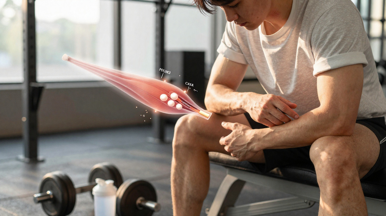
Lifting a sufficiently heavy load causes genuine damage to the muscle fibres responsible for the movement. The minute force-producing units begin to separate at their boundaries, creating small areas of damage within every fibre.
This damage is not evidence that training has gone wrong. What matters is the muscle’s response afterwards: within an hour, its repair system is already in action.
Researchers from three German universities set out to identify the machinery behind that response. They uncovered a set of proteins that detects the damage, labels it and directs it towards disposal.
How muscle damage starts
A sarcomere is the smallest functional component of muscle: a repeating structure that contracts to create force. When millions are arranged end to end, they form a muscle fibre.
When those fibres are put under intense strain through heavy resistance exercise, some sarcomeres collapse. Their normally orderly striped appearance becomes disrupted in localised areas known to researchers as lesions.
These lesions mark the beginning of the process. Before a fibre can restore itself, its damaged components must be removed.
Examining damaged muscle tissue
The research team enrolled eight healthy adults who had not trained their legs for a minimum of four weeks. The group included seven men and one woman.
Every participant completed a demanding leg-training protocol intended to cause muscle damage. It involved single-leg presses, leg extensions and descending 182 stairs while missing every other step.
Thigh muscle biopsies were collected four days before every session and then again 60 minutes after it. Each sample was divided into two portions, allowing the researchers to assess the proteins present in both.
This approach enabled the team to observe muscle during the repair process itself. Only one hour after exercise, markers of damage and proteins involved in repair had already collected in the same sample fraction.
Proteins that react first to muscle damage
A number of proteins rapidly moved towards the injured structures following the workout, including two small heat shock proteins.
PDLIM3, a protein located at anchoring sites within every sarcomere, was another of the early responders.
The researchers followed these proteins after they reached the damaged tissue. They bind to the disrupted structures and pass them into a removal system known as chaperone-assisted selective autophagy, or CASA, as the team calls it.
“Our analytical approaches allowed us to identify proteins that are recruited to the contractile apparatus after resistance exercise, where they perform essential protective and repair functions,” said Professor Pitter Huesgen, a proteomics expert at the University of Freiburg.
CASA allows muscle cells to engulf their broken components and degrade them internally. Removing this debris creates the space needed for repair.
Tracking the clean-up in mouse muscle cells
Although the human samples revealed which proteins accumulated at sites of damage, the team used mouse-derived cultured muscle cells to determine what the individual proteins actually did.
“Using cultured muscle cells, we demonstrated that these repair proteins first recognize damaged muscle structures and then remove them through a cellular degradation pathway known as autophagy,” said Professor Jörg Höhfeld at the University of Bonn.
“This clears the way for muscle repair and adaptation to resistance training.”
Disabling individual proteins in these cells disrupted the entire process. When PDLIM3 was removed, the disposal pathway slowed down and damaged material accumulated.
This suggests that PDLIM3 is an early initiator: one of the proteins that recognises mechanical strain before the clean-up process starts. XIRP1, a marker of muscle damage, was also found to be necessary.
The protective effect fades within weeks
Participants exercised twice weekly for six weeks before taking a three-week break. At every point, the team repeated the muscle-damaging workout and compared what happened.
Following six weeks of training, the identical heavy session produced fewer lesions. The muscles had developed a means of protecting themselves.
Because there was less damage to resolve, the repair proteins showed a less pronounced response. This changed after the three-week pause.
Muscle damage soon rose again to the level observed in untrained muscle. The robust repair response returned alongside it.
Professor Sebastian Gehlert led the human research element at the University of Hildesheim.
“Our findings reveal how training intensity and training history influence both muscle damage and the activation of the repair machinery,” he said.
“This knowledge will help us optimize the sequencing of training sessions for athletes as well as rehabilitation programs for patients in clinical settings.”
Remaining questions
The human component of the study was both small and observational. One woman and one man were excluded, leaving six people in the main analysis, meaning the findings cannot establish differences relating to sex, age or muscle type.
Much of the mechanism itself was identified in mouse cells rather than humans. The authors stress that the human samples establish which proteins travel to damaged sites, whereas the cell studies demonstrate their functions.
The study did not show muscle growth either. Training intensity was low enough that muscle-fibre size remained unchanged, indicating that the protective effect developed without visible growth.
Gehlert is now using these findings in his work training elite athletes at Germany’s Olympic centres and in teaching future exercise scientists.






Comments
No comments yet. Be the first to comment!
Leave a Comment